
Inyan
-
Posts
1,233 -
Joined
-
Last visited
-
Days Won
44
Content Type
Profiles
Forums
Events
Blogs
Gallery
Store
Everything posted by Inyan
-
- 1 reply
-
- 1
-

-
Always my friend
-
Looking at that top slab graft you are trying to do, I can tell a few things... one, you may have used an older possibly a middle cutting, to graft on. Two, dry air or lack of humidity in this case was not your friend this time around. The faster your stock dries out the more of a problem you can have and that is compounded the older your stock is. It is a double edged sword though, too much humidity for too long and you are going to invite rot and infection. Especially so if your tools were not absolutely clean between each cut. This is how I roll with a similar slab graft. Note, the stock in this case was only a few weeks old as it was grafted itself to be used as graft stock...

-
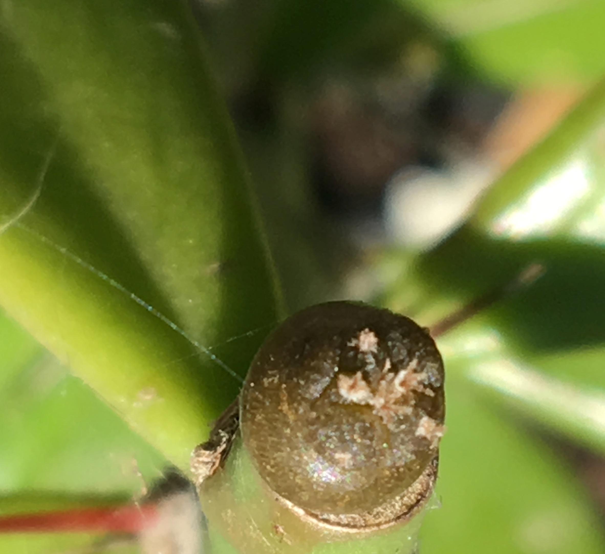
All you need is an areole, but my guess is that one is going to revert to green given enough time.
-
Purple or red... seems to be due to stress from cold or too light... either one can do that.
-
Your Lophophora will not die if you leave the Trichocereus pups intact, but DJ-MC is right... they may steal growth away from your Lophopora scion. So, the question becomes... are you okay with that?
-
 Art is nature or nature is art... either way... that is where I find my peace. It is why I find so much peace in a dab of pollen on that sexy little stigma.
 -
After freezing my collection... down to a very appalling small number now of survivors, I was greeted with signs of flowers today on this little rascal.

-
You should hope for the opposite when testing for cold hardiness the more specimens fail to thrive the more advantage you can see for keeping those that actually made it. I've still got my few frozen bits outside hoping for more freezes, but suffice it to say... I don't see how beneficial that might be seeing that I put thousands of seedlings outside and had less than a handful make it. But, I'm still trying to push the envelope nonetheless.
-
Let's see... I exposed my entire cacti collection to freezing temperatures in an effort to find any that could withstand freezing temperatures. Guess what? 3 Cacti made it! One being a T. grandiflora. Fun times indeed.
-
Looking good Gimli. My favorite way to graft small seedling... especially so if one is making a small army of grafts at the same time is with that parafilm method. Yours is looking good.
-
"'Although all parts of the plant are edible, some reports warn that consumption of large quantities of young shoots can be hallucinogenic and should be avoided." http://www.oardc.ohio-state.edu/weedguide/single_weed.php?id=37 "Although all parts of the plant are edible, some reports warn that consumption of large quantities of young shoots can be hallucinogenic and should be avoided"https://brigittemars.com/other/day-lilies/ Not condoning any lighthearted research with this one, but it does make one want to see what other information is available on the medicinal as well as psychotropic properties of Hemerocallis.
-
Yes, apparently the flowers are edible. The foliage is what is supposed to be hallucinogenic and even then from the little bit I can find on it... it appears that large quantities need to be eaten to garner any affect. If cooked, the foliage becomes edible and the hallucinogenic property is destroyed. I'm thinking juicing might be the best way to gather this substance, but it would be nice to know at what temperature the hallucinogenic molecule is deactivated. Thinking along the lines of drying out the material at highest safe temps to enable a smaller quantity to be worked with. As it is far smarter to err on the side of caution and make many experiments than to overshoot and find out the quantity one has chosen is much more than is safe to work with. "As for edibility….. Young spring shoots and leaves under five inches taste similar to mild onions when fried in butter. They are also a mild pain killer and in large quantities are hallucinogenic. The leaves quickly become fibrous so they can only be eaten young (but you can make cordage out of the older leaves.) The flower buds, a rich source of iron, are distinguished from the plant’s non-edible fruits by their internal layering. The blossoms are edible as well, raw or cooked (as are seeds if you find any.) The dried flower contains about 9.3% protein, 25% fat, 60% carbohydrate, 0.9% ash. It is rich in vitamin A. The closed flower buds and edible pods are good raw in salads or boiled, stir-fried or steamed with other vegetables. The blossoms add sweetness to soups and vegetable dishes and can be stuffed like squash blossoms. Half and fully opened blossoms can be dipped in a light batter and fried tempura style (which by the way was a Portuguese way of cooking introduced to Japan.) Dried daylily petals are an ingredient in many Chinese and Japanese recipes "http://www.eattheweeds.com/daylily-just-cloning-around-2/
-
https://pfaf.org/user/Plant.aspx?LatinName=Hemerocallis+yezoensis " Large quantities of the leaves are said to be hallucinogenic. Blanching the leaves removes this hallucinatory component[205]. (This report does not make clear what it means by blanching, it could be excluding light from the growing shoots or immersing in boiling water[K].)" Anyone know anything about this plant? Is it safe?
-
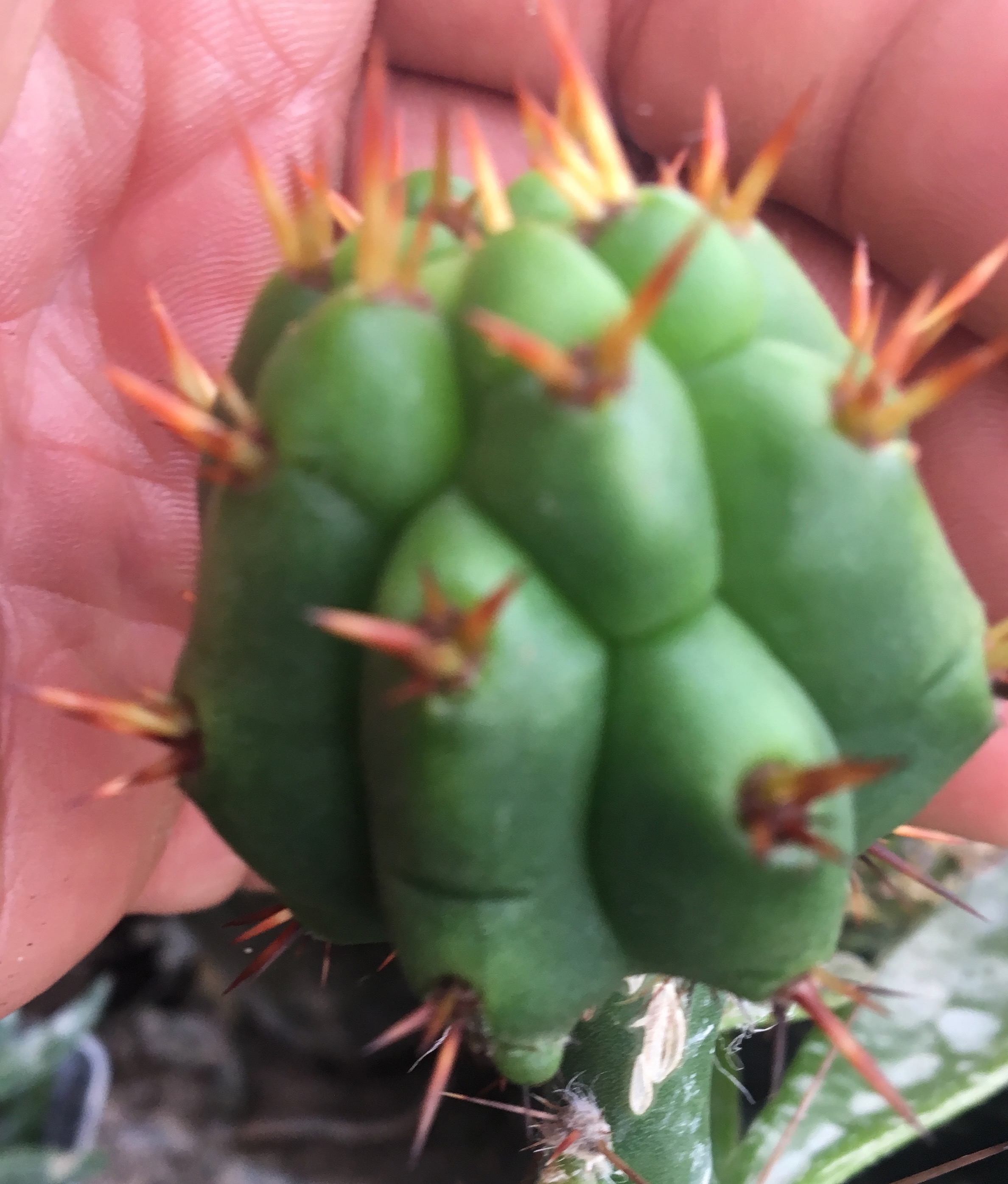
All you need is a single areole on either side and your in business.
-
You never have to put your graft in humidity if your using parafilm. You also never have to remove the parafilm. With that being said, one slice down the side after the graft has taken off should allow the parafilm to be rolled back and easily removed. If its hard to remove your parafilm your trying too hard. You can also simply make your one slice and leave your parafilm in place. The parafilm will fall off on its own. Hope that helps guys. For those that have been wondering what I've been up to... I've subjected my entire collection to freezing weather in hopes of getting a few specimens that can take nice frost, snow, etc. and come up with a very tiny limited gene pool to work with. Very devastating to say the least if I was actually concerned with specimens that lacked hardiness... which I am not. So, here's to having a very few Trichocereus and Echinopsis that made it through the freezing conditions I've exposed them to this year. And when I say a few... I can count my entire collection now on two hands. Ahh, such is life.
-
Pachanoi is most assuredly suited for small seedlings and small areoles to boot....


-
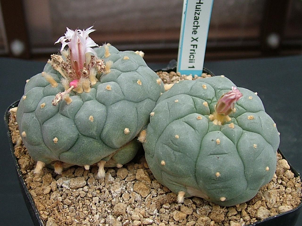
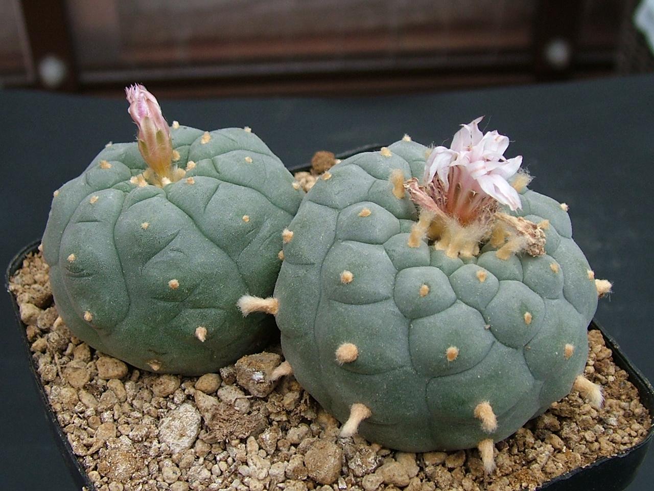
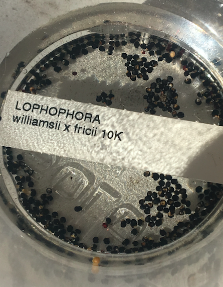
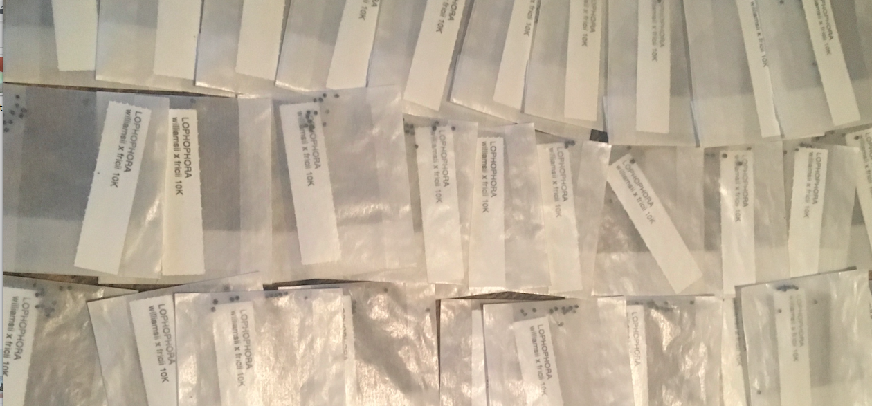
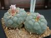
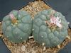
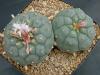
Lophophora w. x L. fricii from KoehresSorry, I was getting impatient waiting for mine to grow.

-
Looks like growth is stalling with lows in the mid 30's and highs in the low to mid 40's at night. Still, I thought I'd share a few pictures so you guys didn't think I forgot about this thread. Just not much to show when its this cold out.


-
Halloween grafting... Halloween grafting.m4v no glasses and just slits to see through. Halloween_grafting.m4v Halloween_grafting.m4v Halloween_grafting.m4v


-
I won't bore you with a long list. Instead, one very high quality cross that I am extremely excited about.... In the cup of water as we speak.

-
-
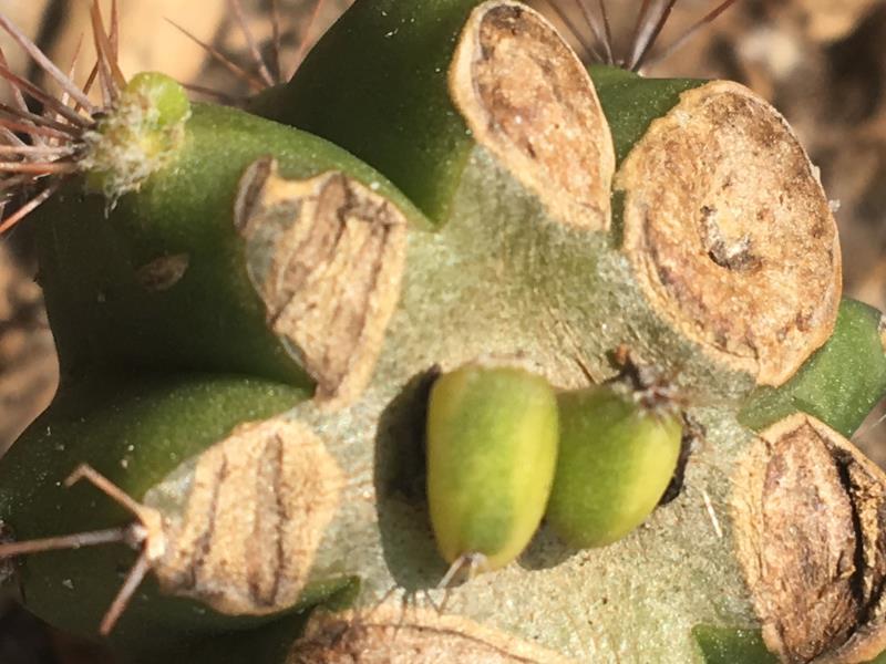
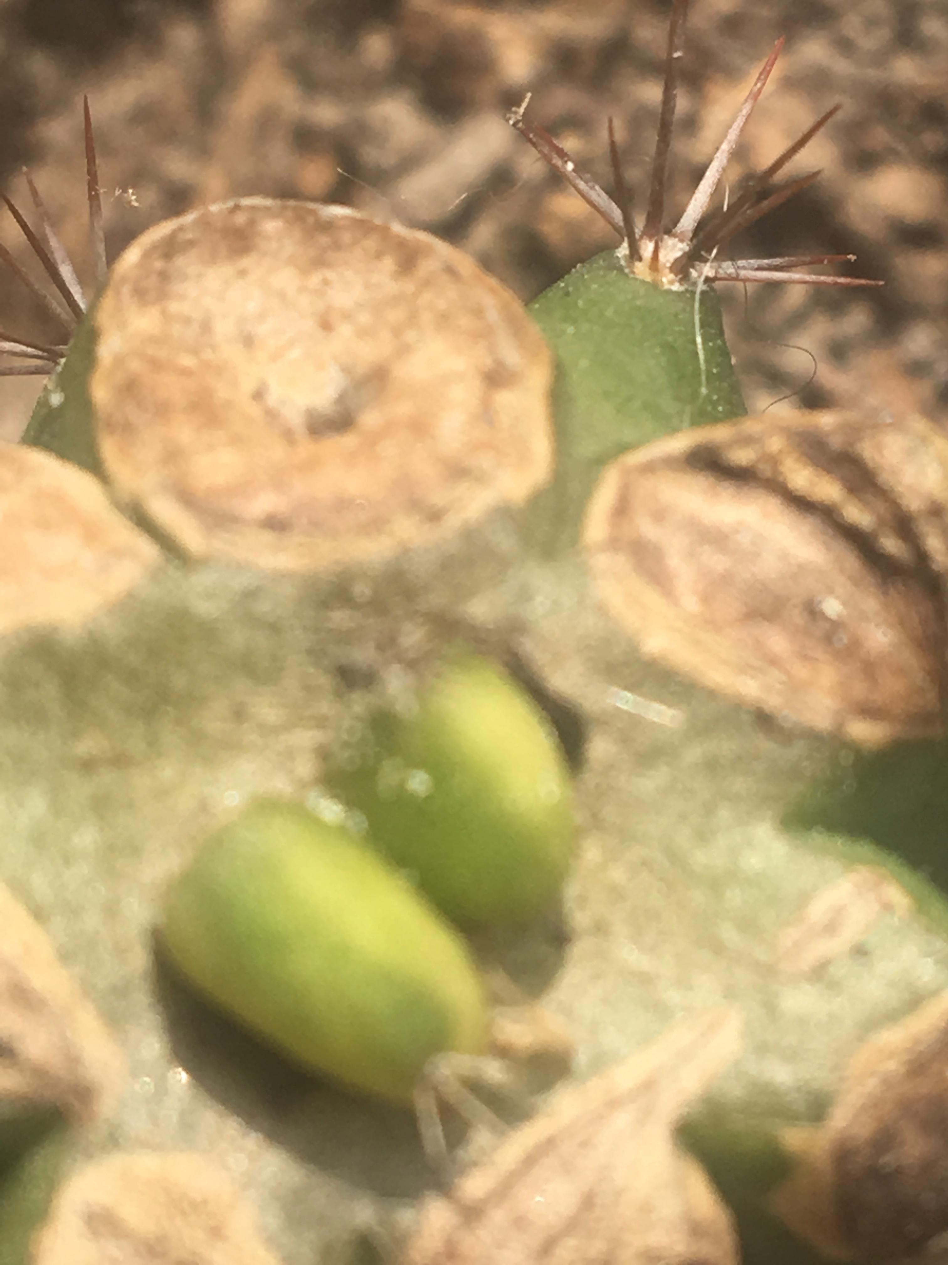
Round two....

-
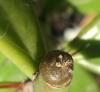
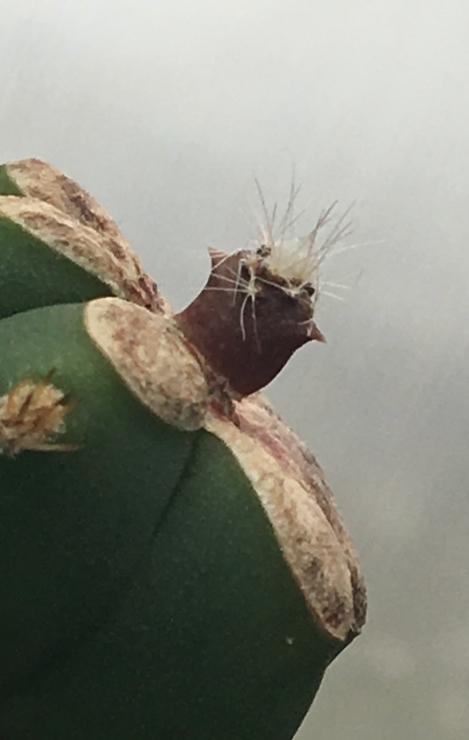
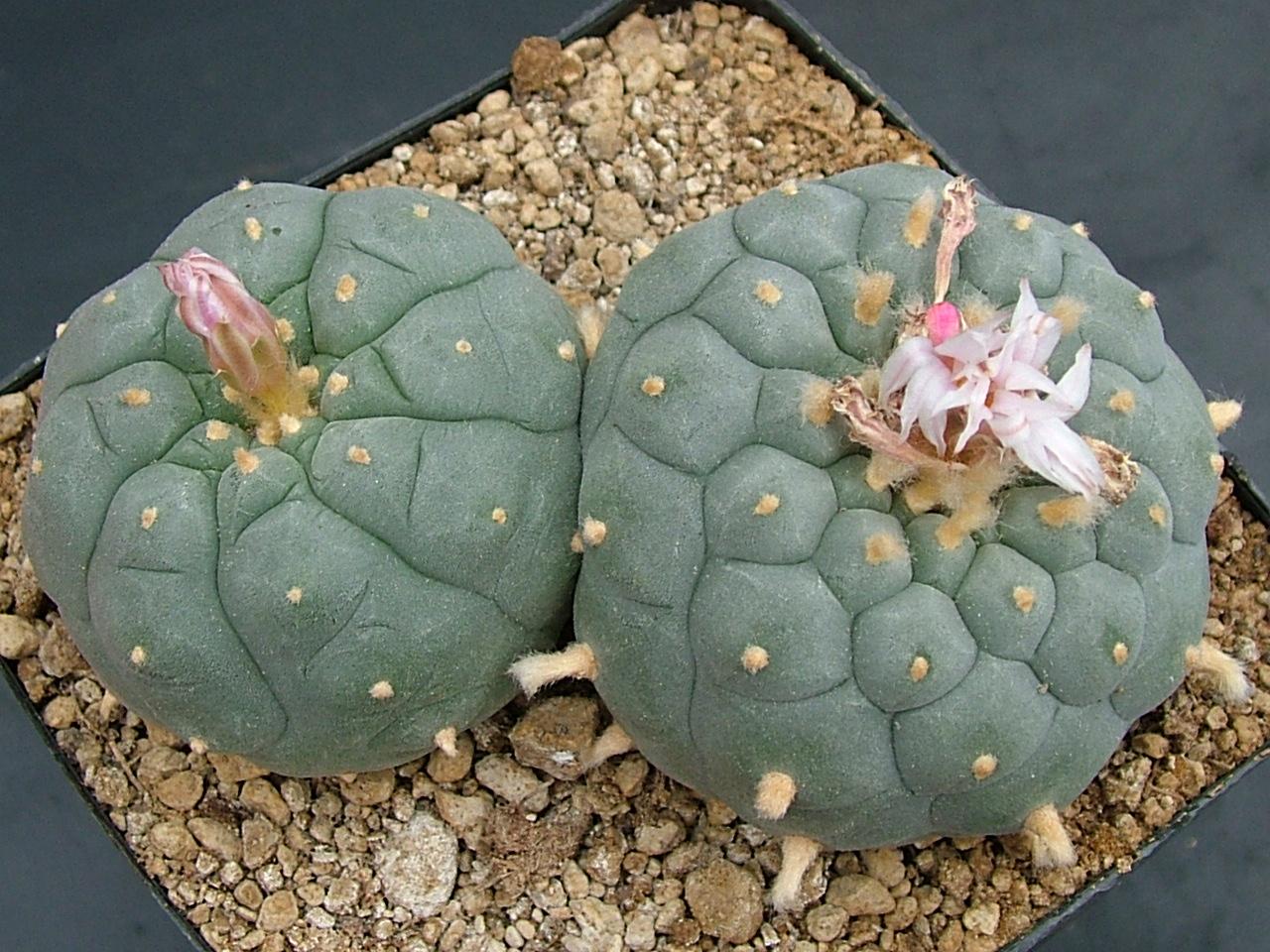
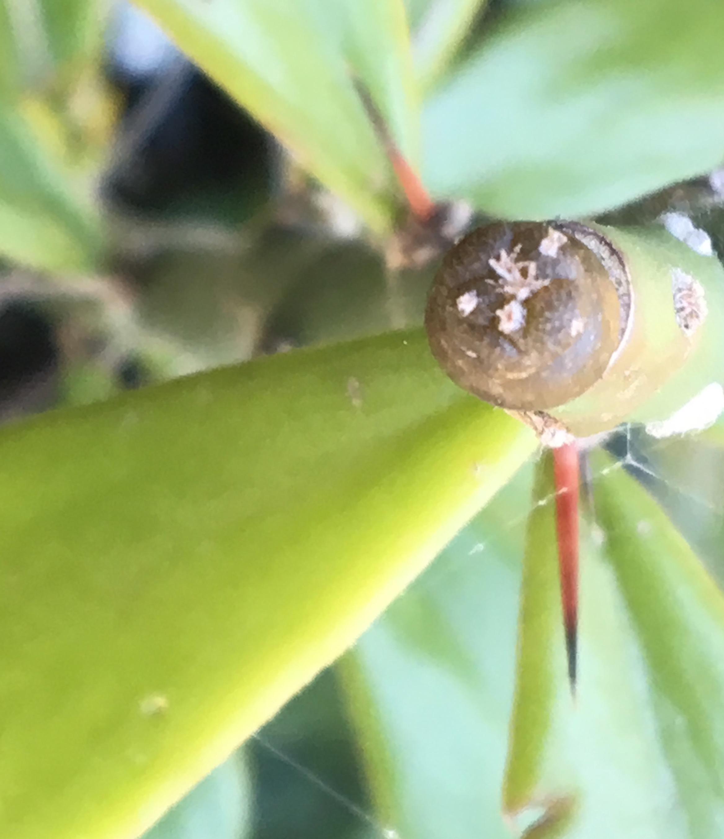
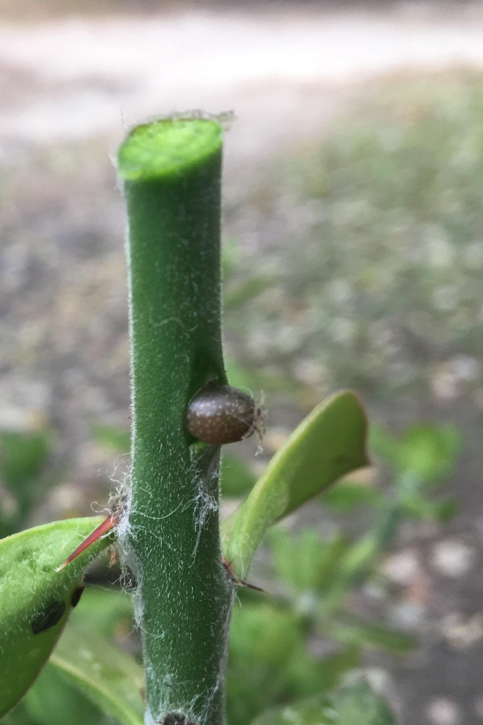
Lophophora williamsii x Lophophora fricii 21 OCT18 26 days